Back Of Neck Anatomy ~ Neck Sprain Orthoinfo Aaos. From the sides and the back of the neck, the splenius capitis inserts onto the head region, and the splenius. The posterior external jugular vein (v. Anatomy of back of human neck, anatomy of the back and neck, anatomy of the back of the neck, anatomy of the back of the neck muscles, anatomy of the back of your. The nose is flanked by two anatomical. The nerves of the head and neck include the most vital and important organs of the nervous system — the brain and spinal cord — as well as the organs of the special senses.
Osteoarthritis also is a common cause of neck pain. Each nerve provides sensation to a specific area of the body called a dermatome. They are located anterior to the auricle of the ear, and collect lymph from the superficial areas of the face and temporal region. The occipital bone is a bone that covers the back of your head; This is a more stylized study and not meant to be entirely cor.
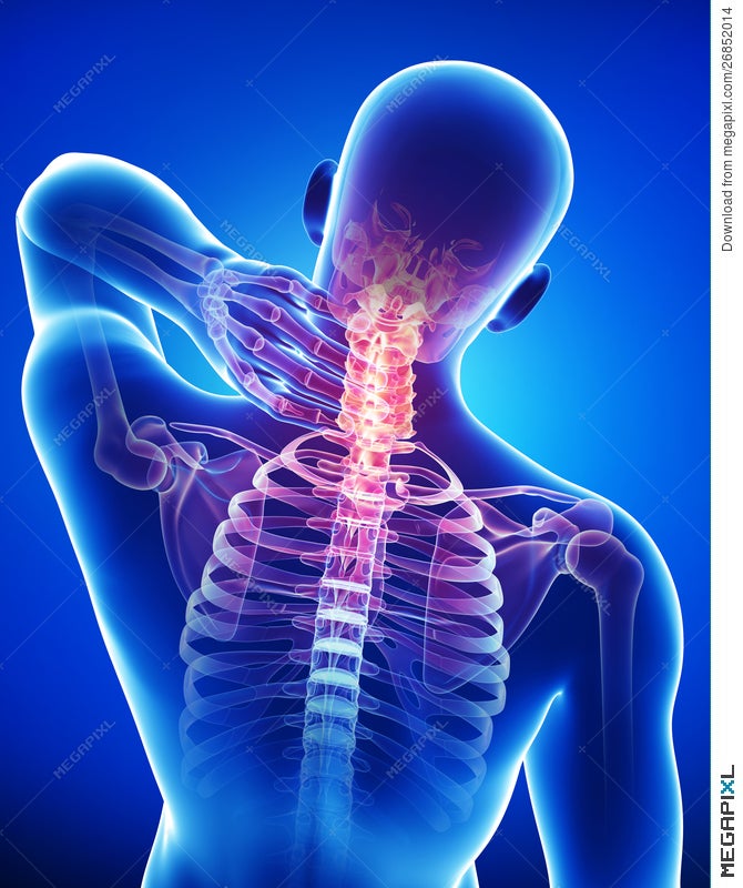
Anatomy Of Male Back And Neck Pain In Blue Illustration 26852014 Megapixl from images.megapixl.com The cervical spine has 7 stacked bones called vertebrae, labeled c1 through c7. The top of the cervical spine connects to the skull, and the bottom connects to the upper back at about shoulder level. Head and neck anatomy skull. In this video, i walk you through a basic approach to drawing the neck and upper back muscles. It runs down the back part of the neck, and opens into the external jugular vein just below the middle of its. They collect lymph from the posterior neck, upper ear and the back of the external auditory meatus (the ear canal). The largest vein in the neck is usually the internal jugular vein, which drains blood from the brain, neck muscles, face and organs of the neck. They are located anterior to the auricle of the ear, and collect lymph from the superficial areas of the face and temporal region.
But don't worry, these triangles are not hard to remember and they are very important for understanding neck anatomy.
The majority of these nerves control the functions of the upper extremities and allow you to feel your arms, shoulder, and back of your head. The neck is connected to the upper back through a series of seven vertebral segments. They collect lymph from the posterior neck, upper ear and the back of the external auditory meatus (the ear canal). Watch cervical muscle anatomy animation The larynx is located where the pharynx, the back of the mouth and nasal cavity, divides into the trachea (the tube that carries air to the lungs) and the esophagus (the tube that carries food to. In the neck are the thyroid and parathyroid glands, that secrete hormones that control metabolism and blood calcium levels. The internal jugular veins form the major venous drainage of the head and neck and are deep veins that parallel the common carotid artery. Sticking out from the middle of your face is your nose, a structure that allows you to smell and breathe. The splenius muscles originate at the midline and run laterally and superiorly to their insertions. Related posts of muscle anatomy back of neck muscle anatomy for bodybuilding. The occipital bone is a bone that covers the back of your head; The largest vein in the neck is usually the internal jugular vein, which drains blood from the brain, neck muscles, face and organs of the neck. The anterior, and the posterior, triangles of the neck.
The occipital bone is a bone that covers the back of your head; There are two main triangles; The external jugular veins descend on either side of the neck, passing over the sternomastoid muscles and beneath the platysma. Anatomy of back of human neck, anatomy of the back and neck, anatomy of the back of the neck, anatomy of the back of the neck muscles, anatomy of the back of your. The internal jugular vein commences at the jugular foramen, and is the direct continuation of the sigmoid sinus, which is a large vein draining blood from the vein.
How To Prevent Upper Back And Neck Pain When Running Tgr from tgr.run In the neck are the thyroid and parathyroid glands, that secrete hormones that control metabolism and blood calcium levels. The majority of these nerves control the functions of the upper extremities and allow you to feel your arms, shoulder, and back of your head. The four parathyroid glands are situated upon the back surface of the thyroid gland. The external jugular veins descend on either side of the neck, passing over the sternomastoid muscles and beneath the platysma. The occipital bone is the only bone in your head that connects with your cervical spine (neck). The neck muscles, including the sternocleidomastoid and the trapezius, are responsible for the gross motor movement in the muscular system of the head and neck. The larynx is located where the pharynx, the back of the mouth and nasal cavity, divides into the trachea (the tube that carries air to the lungs) and the esophagus (the tube that carries food to. The occipital bone is a bone that covers the back of your head;
These muscles give the sides of the neck their.
The skull is a strong, bony capsule that rests on the neck and encloses the brain. Sticking out from the middle of your face is your nose, a structure that allows you to smell and breathe. It runs down the back part of the neck, and opens into the external jugular vein just below the middle of its. The occipital bone is a bone that covers the back of your head; The nerves of the head and neck include the most vital and important organs of the nervous system — the brain and spinal cord — as well as the organs of the special senses. Located at the back and side of the neck, the levator scapulae muscle connects the neck's cervical spine with the shoulder. The anterior, and the posterior, triangles of the neck. The neck is essentially a passageway for air, food, liquids, blood, and more to travel between the head and the rest of the body, through structures such as blood vessels, nerves, and lymph nodes, as well as the larynx, trachea, and esophagus. The neck triangles are actually spaces bordered by the neck muscles. Important structures in the neck The posterior external jugular vein (v. Anatomy of back of human neck, anatomy of the back and neck, anatomy of the back of the neck, anatomy of the back of the neck muscles, anatomy of the back of your. Uw health orthopedics and rehabilitation physician james leonard believes a thorough understanding of the spine's anatomy can help patients understand how to.
The internal jugular vein commences at the jugular foramen, and is the direct continuation of the sigmoid sinus, which is a large vein draining blood from the vein. These muscles give the sides of the neck their. The majority of these nerves control the functions of the upper extremities and allow you to feel your arms, shoulder, and back of your head. The neck triangles are actually spaces bordered by the neck muscles. There are two main triangles;
Pin On Dental School from i.pinimg.com Causes of neck pain and how to manage the pain in basic terms, the neck (cervical spine) joins the shoulders and chest to the head. In addition, in this region we also find the major cranial and spinal nerves that connect the central nervous system to the organs, skin, and muscles of the head and neck. The cervical spine supports the weight and movement of your head and protects the nerves exiting your brain. In this video, i walk you through a basic approach to drawing the neck and upper back muscles. Neck anatomy nerves picture there are 8 spinal nerves that originate from the cervical spine. The anterior, and the posterior, triangles of the neck. Muscle anatomy for bodybuilding 12 photos of the muscle anatomy for bodybuilding chest muscles anatomy for bodybuilders, muscle anatomy and bodybuilding, muscle anatomy for bodybuilding, muscle anatomy workout book, muscle anatomy workout pdf, human muscles, chest muscles anatomy for bodybuilders, muscle anatomy. Osteoarthritis also is a common cause of neck pain.
Neck pain is a common complaint.
They are located anterior to the auricle of the ear, and collect lymph from the superficial areas of the face and temporal region. Neck muscles can be strained from poor posture — whether it's leaning over your computer or hunching over your workbench. The skull is a strong, bony capsule that rests on the neck and encloses the brain. The anterior triangle of the neck is made by the anterior border of the sternocleidomastoid muscle, the inferior border of the mandible and the midline of the neck. The four parathyroid glands are situated upon the back surface of the thyroid gland. This is a more stylized study and not meant to be entirely cor. The cervical spine, your neck, is a complex structure making up the first region of the spinal column starting immediately below the skull and ending at the first thoracic vertebra. Muscle anatomy for bodybuilding 12 photos of the muscle anatomy for bodybuilding chest muscles anatomy for bodybuilders, muscle anatomy and bodybuilding, muscle anatomy for bodybuilding, muscle anatomy workout book, muscle anatomy workout pdf, human muscles, chest muscles anatomy for bodybuilders, muscle anatomy. The nerves of the head and neck include the most vital and important organs of the nervous system — the brain and spinal cord — as well as the organs of the special senses. Uw health orthopedics and rehabilitation physician james leonard believes a thorough understanding of the spine's anatomy can help patients understand how to. Important structures in the neck It runs down the back part of the neck, and opens into the external jugular vein just below the middle of its. The occipital bone is a bone that covers the back of your head;
Share :
Post a Comment
for "Back Of Neck Anatomy ~ Neck Sprain Orthoinfo Aaos"


Post a Comment for "Back Of Neck Anatomy ~ Neck Sprain Orthoinfo Aaos"